Cell biology research and drug discovery demand tools capable of delivering rapid, reliable, and highly detailed results. In this context, PhenoVue™ emerges as a comprehensive solution combining fluorescent dyes, secondary antibodies, optimized microplates, and high-content image analysis systems, all designed to facilitate the most advanced cell assays.
Cell painting and multi-organelle kits: drawing the cellular phenotype
Cell painting is a phenotypic screening technique that provides a complete morphological profile of cells using fluorescent probes that label multiple compartments and organelles. PhenoVue cell painting kits include six probes optimized for staining the nucleus, nucleolus, mitochondria, endoplasmic reticulum, Golgi apparatus, plasma membrane, and actin cytoskeleton. This methodology becomes a powerful tool for studying the effects of chemical compounds, drugs, or genetic modifications.
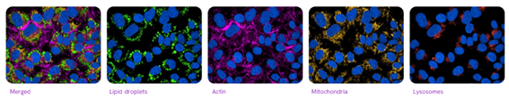
Alternatively, the PhenoVue multi-organelle kit offers a simplified approach to analyzing mitochondria, lysosomes, lipid droplets, actin, and nuclei in fixed cells, ideal for increasing throughput in phenotypic assays without the need to work with live cells.
Specific solutions for neuronal models and microglia
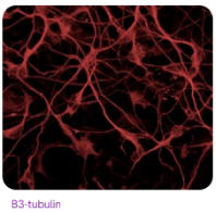
In the field of neuroscience, the PhenoVue line offers researchers staining kits designed to assess neuronal and iPSC-microglia differentiation processes, which are critical in the study of neurological disease models. These kits combine key markers (such as β3-tubulin, nestin, or Iba1) with organelle probes, providing morphological and protein expression data in a single assay.
Beyond the image: cellular functionality and automation
The PhenoVue range is not limited to morphology or structural imaging alone, but also includes reagents for evaluating key functional processes such as viability, apoptosis, mitochondrial membrane potential, intracellular calcium levels, reactive oxygen species (ROS), and hypoxia. This opens the door to more comprehensive experiments in which imaging and function are studied in an integrated manner.
A complete solution: boards, software and automation
This Phenovue range is compatible with the Opera Phenix™ Plus and Operetta CLS™ high-content analysis systems, and with software such as Harmony™ and Signals Image Artist™ , which not only facilitate image acquisition and analysis, but also allow the management of large volumes of data, integration of different types of assays (phenotypic, biochemical, in vivo) and the performance of advanced analyses such as dose-response curves or multivariate classification.
Furthermore, the proposal is complemented by optimized microplates specifically designed for high-content microscopy (PhenoPlate™, CellCarrier™, ViewPlate™) with ultrathin backgrounds, highly transparent materials (such as cyclic olefin foil), or special coatings that facilitate cell culture and/or the formation of 3D spheroids. This ensures high-quality imaging and minimizes common problems such as inaccurate focusing or satellite formation in 3D cultures.
Want to know more?
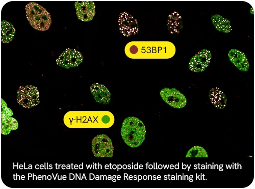
If you would like more information about the PhenoVue™ range presented by Revvity, as well as other cell imaging kits such as the PhenoVue™ DNA Damage Response Staining Kit , their compatibility with different imaging systems, or how they could be integrated into your workflows, our team of specialists will be happy to advise you . Contact us and tell us about your needs: we will help you find the most suitable solution for your laboratory.